Please note that these notes are in progress of being edited!
(1) Define resolution and magnification.
(2) Explain the differences between these terms (with reference to light microscopy and electron microscopy).
Microscopy
Light and electron microscopy
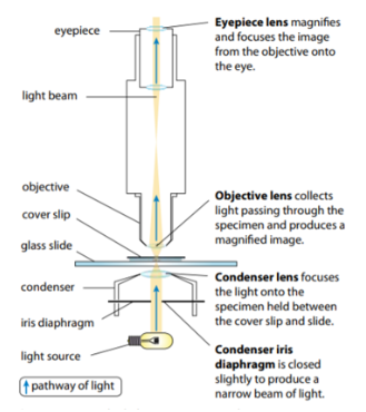
- There are two types of microscopes — light microscope and the electron microscope.
-
- Both use a form of radiation (light/electrons) to create an image of the specimen.
- Light microscopes use an objective lens and eyepiece lens to focus light rays.
-
- The radiation passes through the specimen on a slide and is focused by the lenses.
- The objective lens and eyepiece lens both act to focus the radiation.
- This produces a magnified image of the specimen on the retina of your eye.
- An electron microscope uses beams of electrons rather than light rays.
-
- The specimen must be thin and placed in a vacuum to allow electrons through.
- The electrons are focused onto a screen (where they form a magnified image).
- Electron microscopes offer significantly higher resolution than light microscopes.
- There are two types of electron microscopes (TEM, SEM).
-
- Transmission electron microscopes (TEM) pass electrons through the specimen before being viewed.
-
-
- Only the electrons that are transmitted (passed through the specimen) are seen.
- This allows us to see thin sections of specimens (and thus to see inside cells).
-
-
- Scanning electron microscopes (SEM) uses the electrons to scan the surfaces of structures with only the reflected beam being observed.
-
-
- SEMs allow surface structures to be seen (in contrast to TEMs).
- SEMs also achieve great depth of field — much of the specimen is in focus at the same time, achieving a 3D appearance.
- SEMs do have lower resolutions (3-20 nm) compared to TEMs (0.5 nm).
-
(3) Calculate magnifications of images and actual sizes of specimens from drawings, photomicrographs and electron micrographs.
Magnification
Formula
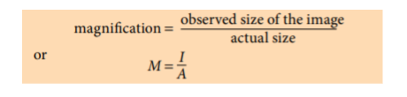
- Magnification is the number of times an image is larger than the real size of the object.
-
- To calculate ‘M’, divide the size of the image by the size of the actual object.
- ‘I’ is the observed image size (what you measure with a ruler).
- ‘A’ is the actual size of the object (size of the cell before it is magnified).
- Before calculating, convert every measurement to µm.
- There is no limit to the extent to which you can magnify an image.
- The amount of useful magnification depends on microscope resolution.
Calculations with a given magnification
- The magnification is given as x100,000 (‘M’ in the formula).
- Measure the length of the image with a ruler (‘I’ in the formula).
- Convert the measurement to µm by multiplying by 10000.
- Substitute ‘M’ and ‘I’ into the formula to get ‘A’.
Calculations with no given magnification
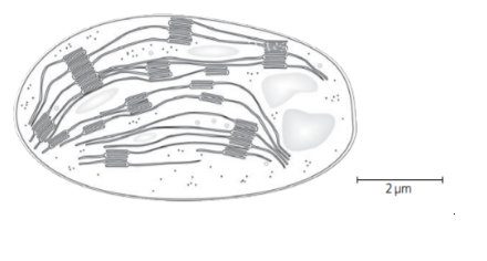
- Measure the length of the scale bar (‘I’ in the formula).
- Convert the measurement to µm by multiplying by 10000.
- The number written at the scale bar is the actual size (‘A’ in the formula).
- Calculate the magnification using the formula.
- Measure the length of the image and convert it to µm (‘I’ in the formula).
- Calculate its actual length using the formula (you have both ‘M’ and ‘I’).
PDF Loading…